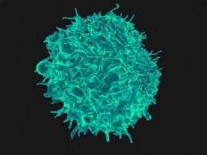
A colorized scanning electron micrograph of a T cell from NIAID.
Next generation therapies and vaccine research have recently dominated headlines and scientific discovery. This research and its ability to treat previously untreatable conditions has rightfully excited the scientific and medical communities worldwide. But the complexities involved require a nuanced approach to ensure the treatment is safe and effective. Integrating cell culture into cellular immunity testing can be challenging, which is why the bioanalytical community has sought options beyond traditional immunoassays. Enter ELISpot assays.
ELISpot assays boast unprecedented sensitivity and specificity. They could legitimately shape the future of cellular immunogenicity testing. As scientists push the boundaries of drug discovery and development, they will inevitably turn to the best tools available. ELISpot assays, with their unique capabilities, will prove to be indispensable companions in that journey.
How ELISpot assays work
One of the primary challenges in understanding cellular immunity is the presence of naïve T cells that do not produce cytokines. But when an infection, natural exposure or immunization occurs, the response happens within these T cells. They multiply and are trained to undertake effector functions. Then, when these memory T cells re-encounter an antigen, they secrete the specific cytokines they were programmed to produce.
The genius of ELISpot assays lies in their ability to provide a visual representation of the secretion from individual cells, which helps determine the frequency of cytokine production. This technique offers the highest sensitivity and efficiency for direct in vitro detection of antigen-specific T cells. In fact, the assays provide ex vivo frequency measurements with exceptional sensitivity, reaching down to the 1/1,000,000 cell range. This can be a game-changer in reflecting the magnitude of T cell immunity, determining the quality of T cell immunity, and even identifying CD4 or CD8 T cell antigen-specific determinants.
Affinity determination is another advantage in determining the affinity of antigen-specific T cells. And unlike many cell detection techniques, ELISpot requires remarkably small sample sizes. But ELISpot assays aren’t without limitations.
Their level of precision is significantly influenced by the detection antibodies and reagents used. The antibodies must be carefully matched to the cytokine under observation, as cells could secrete numerous cytokines and other proteins that might interfere with accurate detection. This could result in high background coloration, making distinguishing and counting spots difficult. Moreover, while isolating PBMCs from whole blood for analysis, the unique characteristics of every serum batch can impact T cell performance in vitro, emphasizing the critical role of serum in both PBMC freezing and ELISpot testing.
Deciphering ELISpot data
The true power of ELISpot lies in data acquisition and the effective interpretation of this data. Various metrics derived from ELISpot data serve as signposts guiding researchers toward insightful conclusions and informed decision-making.
First, spot count stands as one of the most fundamental metrics. Every spot on an assay plate represents an individual immune cell secreting the target molecule, whether a cytokine or another bioactive agent. As researchers tally these spots, they’re effectively gauging the magnitude of the immune response. A surge in spot count typically signals an intensified immune engagement, making it a valuable marker when comparing immune responses across different groups or conditions.
Complementing spot count is spot size. Much like a magnifying glass revealing finer details, the size of each spot explains nuances about target molecules’ secretion levels. Essentially, larger spots paint a picture of more copious secretion by immune cells, offering an additional layer of depth to understand the quality of immune responses.
Furthermore, spot intensity measures the vigor with which each spot shines. This metric acts as a barometer for the strength of the immune response. An uptick in spot intensity typically resonates with a more robust secretion of the target molecule.
Background subtraction also plays a vital role in ensuring data accuracy. By neutralizing the background noise—i.e., signals not derived from specific immune responses—this process ensures that the spotlight remains solely on the specific immune interactions of interest.
Finally, spot-forming units (SFU) showcase the assay’s adaptability, accounting for variables like dilutions or the specific area under analysis. By harmonizing these factors, SFU provides a standardized platform for comparing immune responses across different samples or conditions. This standardization empowers researchers to draw parallels, identify outliers and make informed decisions.
ELISpot vs. alternative methods
Given its sensitivity and specificity, ELISpot has firmly established its position in cellular immunogenicity testing. However, the true measure of its strengths and limitations is best understood by comparing the assays to alternative methods. Here, we look at ELISA, flow cytometry, bead arrays and sample constraints.
- ELISA is adept at detecting the concentration of cytokines produced by cells but does not offer insights into the frequency or magnitude of cells producing these cytokines. Moreover, while ELISpot provides information about the number of cells secreting a particular cytokine, ELISA gives quantitative data about the absolute amount of cytokine present in a sample.
- Flow cytometry
-
- Phenotyping: Flow cytometry excels in scenarios requiring detailed phenotypic information about the cytokine-producing cells. For instance, when there’s a need to discern which specific T cell subtype produces the cytokine, flow cytometry becomes indispensable.
- Intracellular analysis: Flow cytometry remains the method of choice for intracellular cytokine staining to understand cytokines stored within a cell but not yet secreted.
- Kinetic studies: Unlike the static snapshot provided by ELISpot, flow cytometry can be employed for real-time kinetic studies, tracing how cytokine production evolves.
- Live cell sorting: When the goal is to segregate live, cytokine-producing cells for subsequent culture or analysis, technologies like fluorescence-activated cell sorting (FACS) offer clear advantages.
- Multiplex or cytometric bead arrays (CBAs): These techniques are most appropriate when research requires simultaneous multiparametric analysis of several cytokines. ELISpot, by design, is usually optimized for single-cytokine detection.
- Sample Constraints: One of ELISpot’s most significant advantages is requiring fewer cells than other techniques, but exceedingly limited samples—i.e., those procured from certain biopsies—could still pose challenges.
While ELISpot assays offer high sensitivity and efficiency in specific cellular immunogenicity contexts, alternative methods provide unique advantages depending on the study’s objectives and constraints. The specific goals of the study should always guide the choice of the method, and each tool’s strengths and limitations should be considered.
ELISpot assays: Preclinical discoveries to clinical trials
the ELISpot assay has emerged as a particularly robust and reliable tool within the diverse toolkit available to scientists during preclinical investigations. In the vast landscape of immune response assessment, ELISpot assays stand out in their capacity to enumerate antigen-specific T cells actively producing pivotal cytokines like interferon-gamma (IFN-γ) or interleukin-2 (IL-2). As potential vaccine candidates are evaluated, ELISpot assays assist researchers in gauging the efficacy of these vaccines by quantitatively measuring the immune responses they invoke.
Furthermore, these assays are adept at managing the intricate interaction between immune cell activation and differentiation. For example, when peeling back the layers of T helper subsets like Th1, Th2, and Th17 or the workings of cytotoxic T cells, ELISpot proves indispensable. Such insights facilitate a richer characterization of immune cell populations and their associated functional traits.
Finally, ELISpot assays are at the forefront of screening and characterizing promising immunotherapies. Whether they are vaccines or novel immunomodulatory agents, ELISpot helps researchers pinpoint drugs that evoke the desired immune responses and/or focus on specific cellular targets. Moreover, it holds keys to unlocking deeper understandings of immune tolerance and regulatory mechanisms, especially when studying the roles and functionality of regulatory T cells (Tregs) or other immunosuppressive cell cohorts.
Investigational New Drug (IND)-enabling studies ensure that only the most promising candidates progress to clinical trials. ELISpot assays can play a pivotal role in this endeavor.
By quantifying cytokine-producing stalwarts of the immune system—i.e., T cells, B cells and NK cells—ELISpot assays paint a comprehensive picture of a drug’s interaction with the immune system. Moreover, its versatility extends to the pharmacodynamics of drug candidates. As these potential drugs engage with the body’s immune machinery, ELISpot assays track real-time cellular responses—from cytokine production to cytotoxicity. Such insights strengthen the scientific understanding of the drug’s mode of action (MOA) but also arm researchers with the empirical evidence needed to make informed decisions about advancing these candidates to clinical testing.
Key takeaways for drug developers and sponsors
For drug developers and sponsors, there are two undeniable truths about ELISpot assays. First, they offer significant scientific value in their sensitivity and specificity. By capturing the whispers of cytokines secreted by individual immune cells, ELISpot assays serve as a barometer for understanding the nuanced immune modulations induced by potential drugs.
Second, ELISpot assays hold a special place in evaluating vaccine candidates. By focusing on the chorus of cytokines from immune cells, ELISpot assays offer insights into a vaccine’s protective shield against pathogens. This knowledge isn’t just academic—it’s the foundation for drug developers and sponsors to build effective vaccine candidates.
The bottom line on ELISpot assays is that their multifaceted applications make them indispensable allies in any drug candidate’s journey. The data they provide promises a safe, innovative and effective approach to future drug development.
About the authors
Dan Wang, MS: Dan is Immunology, Group Leader/Study Director/Principal Investigator/Contribution Scientist at WuXi AppTec. A distinguished immunologist with a robust background in applied chemistry, Dan has been with WuXi AppTec since 2018. His academic journey began with a bachelor’s degree in applied chemistry from Anhui Normal University and a master’s degree in chemistry from Southeast University.
Dan is proficient in ELISA and other immunological technologies, adept with analytical software and committed to good laboratory practices. He has also completed extensive internal training in Good Documentation Practice (GDP), Good Laboratory Practice (GLP), personal protective equipment (PPE), and animal welfare and bio-chemical safety protocols.
Tingting Yu, BS: Tingting is Immunology, Group Leader/Study Director/Principal Investigator/Contribution Scientist at WuXi AppTec. A resourceful and skilled immunologist, Tingting holds a bachelor’s degree in biological pharmaceuticals from Dalian Medical University. She has been a group leader in the Immunology Anti-Drug Antibody Group since August 2018.
Tingting’s roles at WuXi AppTec have included assistant director, study director, principal investigator and contributing scientist. Tingting is also responsible for developing new immunology assays and maintaining laboratory equipment.
Dan Hu, MS:
Dan is Group leader, Immunochemistry Principal Investigator/Study Director and Project Manager, overseeing the immunochemistry team within the bioanalytical services (BAS) department at WuXi AppTec. He is an experienced bioengineer with a master’s degree in biological engineering from the East China. University of Science and Technology, where she also completed an undergraduate degree with a minor in English.
Filed Under: Biologics, Cell & gene therapy, clinical trials, Drug Discovery, Drug Discovery and Development, Genomics/Proteomics, Immunology


