[Image courtesy of Thermo Fisher Scientific]
Spatial biology involves the use of advanced imaging and molecular techniques to visualize and map the location of cells, transcripts, proteins and their interactions within tissues. Traditional biological studies have long focused on analyzing individual components of cells in isolation. Spatial biology analyzes intact tissues in space, offering a more accurate and holistic view of biological processes. These approaches often overlook spatial relationships and contextual interactions that are crucial for a comprehensive understanding of cellular function. Spatial biology addresses this gap by emphasizing the importance of spatial context in studying cellular environments.
Driving progress in cancer research and precision medicine
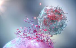
One of the most promising applications of spatial biology lies in the realm of cancer research. Scientists can map the spatial organization of cells within tumors and identify how cancer cells interact with their surrounding microenvironment, including immune cells and blood vessels. This knowledge can reveal new therapeutic targets and strategies to disrupt these interactions, thereby inhibiting tumor growth and metastasis. In the neuroscience field, spatial biology allows for detailed mapping of brain tissue which reveals the intricate networks of neurons and glial cells. Understanding these connections is vital for unraveling the complexities of Alzheimer’s and Parkinson’s disease, epilepsy and neurodevelopment disorders.

[Image courtesy of Thermo Fisher Scientific]
These technologies will transform personalized medicine as they become more accessible and integrated into clinical workflows. Physicians will be able to make more informed decisions based on the spatial context of molecular data, leading to better patient outcomes and more efficient healthcare. Ultimately, the integration of spatial biology into clinical practice promises to revolutionize the way we diagnose, treat and understand complex diseases. We can expect significant advancements in diagnostics, therapeutics and overall patient care as we continue to uncover the spatial dynamics of cells.
Spatial proteomics, a subset of spatial biology, plays a crucial role in this transformation. By leveraging advanced imaging techniques and sophisticated computational tools, spatial proteomics enables scientists to visualize and analyze the spatial organization of proteins within cells and tissues with incredible precision. This approach supports the mapping of protein interactions and distributions which reveals how proteins influence cellular functions and disease mechanisms.
This method of research supports cancer research by helping scientists study the spatial organization of cells within tumors. It can also help map the intricate networks of brain cells in neuroscience by aiding in the development of therapies for neurological disorders.
Making spatial scalable
Spatial proteomics is at a pivotal stage, allowing a broader spectrum of scientists to extract more insights from their tissue samples without incurring additional costs or complexity. However, key challenges remain in making these technologies more accessible and user-friendly, including the complexity of data analysis, high costs, labor-intensive sample preparation and the need for specialized technical expertise.
Now, there are new innovations that are helping to address these issues through breakthroughs in software development within the microscopy system, simplifying image processing and data interpretation. Additionally, the industry is adopting and energizing existing laboratory technologies to streamline sample preparation and staining processes.
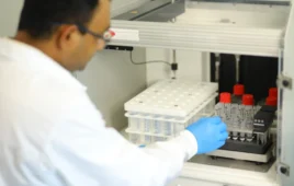
Recognizing the high costs of sophisticated equipment and reagents, Thermo Fisher recently introduced the Invitrogen EVOS S1000 Spatial Imaging System. Innovative products like this can help overcome the limitations of current fluorescent microscopy technologies, enabling researchers to generate multiplexed, high-quality images for multiple samples within hours, thereby lowering the barrier to entry into spatial tissue proteomics.
Higher resolutions and techniques such as multiplexed imaging provide intricate maps of cellular architecture within tissues. In parallel, the integration of advanced data analysis methods, including machine learning and artificial intelligence, has transformed the way we interpret complex biological data. These tools can process vast amounts of information quickly and accurately, identifying patterns and insights that would be impossible to discern manually.
Efforts like this are crucial for democratizing access to spatial biology, thus empowering a larger scientific community to leverage these advanced tools in their research.
Spatial proteomics represents a paradigm shift in the way we study and understand biological systems. By embracing the spatial dimension, we can uncover new layers of complexity and gain insights that were previously beyond our reach. This field’s continuous evolution holds the promise of transforming research and clinical practice, ultimately leading to a new era of precision medicine where treatments are tailored to the unique spatial context of each patient’s disease.

Trisha Dowling
About Trisha Dowling
Trisha Dowling currently serves as a vice president at Thermo Fisher Scientific, where she specializes in cellular analysis and instrumentation. She has more than 25 years of experience in life sciences industries like biotechnology and pharmaceuticals, including an extensive background in marketing, product development and business management.
Filed Under: Biospecimens, Biotech, Genomics/Proteomics