[Adobe Stock]
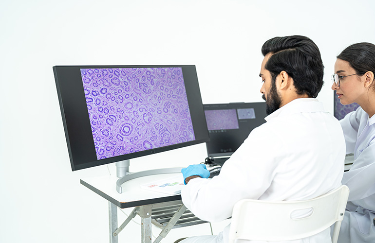
Traditionally, the default path of slides was something like a one-way street. A pathologist read a slide, wrote down the main findings and, after that, the glass went into storage. “Before, the pathology endpoint was just a report,” said Aleksandra Zuraw, DVM, PhD, a veterinary pathologist at Charles River Laboratories. “Now you have the digitized slide, which is pixels.”
Researchers can pair those images with pathology reports and molecular measurements to train models to recognize patterns associated with specific biological changes. A slide can then become a source of data for questions beyond the original pathology assessment.
Researchers can also return to the tissue used to make those slides. Molecular tests on stored samples may help explain how a drug affected cells and organs, answering questions that emerged after the original animal study ended.
Getting more from the animals a study already uses
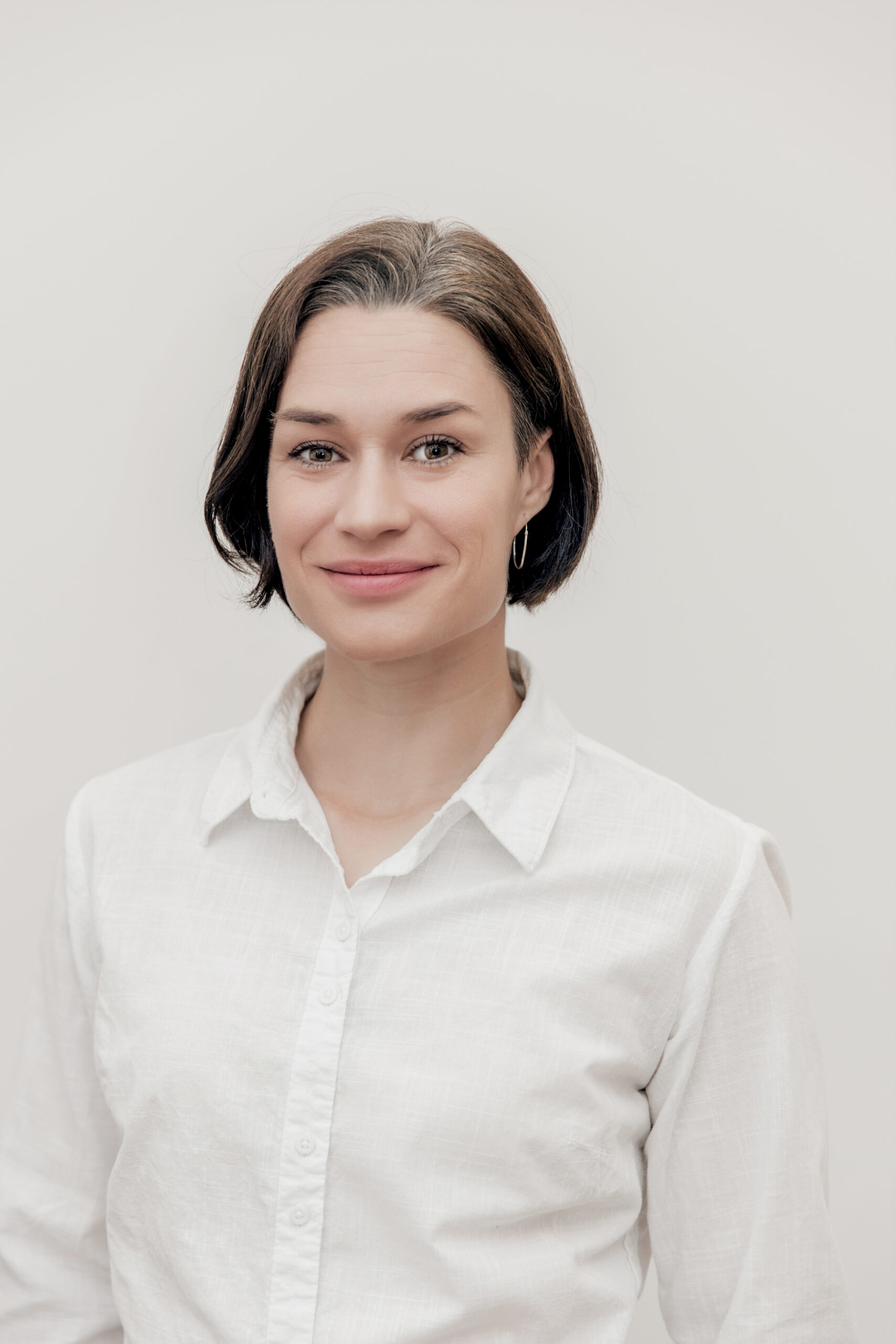
Aleksandra Zuraw, DVM, PhD
Charles River, which is one of the largest global companies specializing in preclinical animal research and experimental animal models, launched its Alternative Methods Advancement Project in April 2024.
One of the company’s shorter-term initiatives in this transition is to collect more from the animals a study already uses. One route is virtual control groups. In a standard toxicology study, each dose group is paired with a concurrent control group that is handled identically but doesn’t receive the drug. “Control animals are always a big fraction of every study,” Zuraw said, so replacing some of them with matched historical data cuts animal numbers by simple arithmetic.
Using historical controls requires researchers to establish that the earlier animals and study conditions are comparable enough to provide a reliable baseline. Otherwise, differences between studies could be mistaken for effects of the drug. Zuraw said Charles River’s virtual control group team is working on “generating enough matched data to provide virtual controls wherever it’s possible to provide them.”
Archived tissue that keeps its study context
In toxicology studies, researchers examine tissue for signs that an experimental drug has damaged organs or cells. They typically preserve samples in formalin, a formaldehyde solution, and embed them in paraffin wax before cutting thin sections for slides. The remaining formalin-fixed, paraffin-embedded (FFPE) blocks go into an archive.
Those blocks can retain molecular information about how the tissue responded to treatment. Collecting fresh-frozen tissue for molecular analysis generally requires planning before a study begins. When a question arises afterward and suitable samples are unavailable, researchers may need another animal experiment, Zuraw said.
On her podcast, Zuraw talks with Syed T. Hoda, M.D., director of digital pathology at NYU, about moving a department of roughly 95 pathologists to digital sign-out in a single push. [Dr. Aleks Digital Pathology & AI/YouTube]
The Organisation for Economic Co-operation and Development’s 2025 guidance on sample collection for omics analysis describes how preserved tissue can support molecular investigations, including studies of gene expression and proteins. It also stresses that sample preparation and storage affect the quality of the results.
“So now you have the option to use FFPE material that was already part of a lot of studies for additional information,” Zuraw said. “You don’t lose the context of the study.”
Researchers can compare the additional molecular findings with the original dosing information, tissue observations and control data. As Zuraw put it, they can “extract more from the same animals, from the same blocks, without running a new experiment.”
Using archived samples to avoid additional animal experiments fits into a broader regulatory push starting with the FDA Modernization Act 2.0, enacted in 2022, which allowed nonanimal methods to support applications to begin human drug trials. Since then, the agency has provided more concrete guidance, including updates in September 2026. In April 2025, FDA published a roadmap for reducing animal testing in preclinical safety studies, starting with monoclonal antibodies and later expanding to other biologics and eventually new chemical entities. Over three to five years, the agency aims to make animal studies the exception rather than the norm, with new approach methodologies (NAMs) as the default.
New methods for data sources that already exist
Zuraw points to a series of parallel developments impacting the field, including molecular prediction from hematoxylin and eosin (H&E)-stained slides, an approach whose use is growing in medical diagnosis and research. She refers to it as “a big area of development.” She explains: “You take a data source, a digitized H&E image, you don’t even have to use other methodologies, and you apply new methods. Whatever those methods turn out to be, if you know the principle, you know they can be applied to a digital slide.”
Virtual staining, for instance, is expanding quickly, which Zuraw notes potentially involves “skipping the staining and skipping glass slides altogether,” and is “more cutting-edge.” One 2026 review documents an accelerating proliferation of applications across tissue types, tracing back to demonstrations in 2019.
Most virtual staining still starts with a glass slide: a scanner captures an unstained tissue section, and software generates the appearance of a stain. Zuraw sees a wider cycle taking shape. As researchers find new uses for digital images, labs have more reason to invest in the systems needed to create and share them. “I hope it becomes a snowball effect,” she said, opening up “data sources that aren’t new, but that we didn’t have a way to access before.”
Turning regulatory guidance into routine practice will require labs to demonstrate how these methods work in actual studies. “You still need enough early adopters to generate precedent,” Zuraw said. “Those early adopters will embrace the guidance, figure it out and troubleshoot, and then others can build on their work without reinventing the wheel.”
Filed Under: Drug Discovery