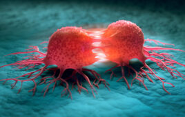
A study led by a team of Massachusetts General Hospital (MGH) investigators has analyzed, for the first time, the mechanisms underlying the use of focused ultrasound to improve the delivery of anti-cancer drugs across the blood brain barrier into brain tumors. Their report published in PNAS uses advanced microcopy techniques and mathematical modeling to track the potential of this promising, minimally invasive treatment approach in an animal model of breast cancer brain metastasis. The team also included investigators from Georgia Institute of Technology, the University of Edinburgh and Brigham and Women’s Hospital.
“The blood brain barrier is a challenge in the treatment of brain malignancies, as it can hinder drug delivery,” says co-corresponding and co-lead author Costas Arvanitis. “Even when a drug reaches the brain’s circulation, abnormal blood vessels in and around tumors lead to non-uniform drug delivery, with low concentrations in some areas of the tumor. If a drug makes it to a region of the tumor and crosses the abnormal blood vessel wall, it encounters dense tissue within the tumor that can block access to malignant cells. We sought to use a new methodology that may improve these abnormal transport properties to enhance drug delivery and efficacy throughout a brain tumor.” Arvanitis, an assistant professor at Georgia Institute of Technology, worked on this study at both the Edwin L. Steele Laboratories for Tumor Biology in the MGH Department of Radiation Oncology and in his new Biomedical Acoustics and Image-Guided Therapy laboratory at Georgia Tech.
Focused ultrasound concentrates multiple beams of ultrasound energy on a single spot within the body. Microbubbles – tiny lipid bubbles that vibrate in response to ultrasound signals – injected into the circulation can temporarily breach the blood brain barrier at the target site. Although this approach has been studied in animal models with promising results – leading to phase 1 clinical trials in conditions including primary brain tumors such as glioblastoma – the underlying mechanisms have not been well understood. To learn more about the properties of focused ultrasound treatment of brain tumors, the team employed advanced microscopy techniques in live mice that had received implants of HER2-positive breast cancer cells in their brains.
In their experiments, the researchers explored the ability of focused ultrasound to enhance delivery of two types of anti-cancer agents – the common chemotherapy drug doxorubicin and the targeted drug T-DM1, which combines the HER2-antibody-based drug trastuzumab (Herceptin) with cytotoxic DM1. Not only did the approach improve delivery of both drugs across blood vessel walls, with even greater improvement for the smaller doxorubicin molecule, it also improved the distribution of both drugs within tumor tissue. For the first time, the MGH team’s experiments showed that focused ultrasound enhanced the permeability of the endothelial cells that line tumor blood vessels, leading those cells to take up doxorubicin.
“Evidence of increased cellular transmembrane transport and uptake of doxorubicin by focused ultrasound was largely unknown until now,” says co-lead author Vasileios Askoxylakis, MD, of the Steele Labs at MGH. “And while focused ultrasound with microbubbles also increased the penetration of the antibody-drug conjugate T-DM1 into tumors, that improvement diminished five days after drug administration, supporting the hypothesis that T-DM1 accumulation is a result of transiently increased permeability of tumor blood vessels.”
High-resolution imaging allowed the investigators to demonstrate increased flow of the interstitial fluid between cells within a tumor after the application of ultrasound and to reveal its role in improving drug delivery. Mathematical modeling enabled the investigators to quantify focused-ultrasound-induced changes in tissues and cellular transport properties and to identify optimal conditions for improved drug delivery. These results could provide a framework for the development of new strategies and the design of clinical trials that combine promising therapeutics with focused ultrasound.
“By explaining and underscoring the potential of combining focused ultrasound with different drugs for the treatment of brain metastases, our findings provide important scientific principles for the optimal clinical use of the technology,” says senior author Rakesh Jain, director of the Steele Labs for Tumor Biology and the Cook Professor of Radiation Oncology at Harvard Medical School. “In particular, they may allow identification of specific administration protocols for improved drug uptake – such as slow infusion rather than bolus administration – and support the hypothesis that the approach needs to be tested individually for different drugs. By laying the groundwork for more rational design and deeper understanding of focused-ultrasound-based treatment, our work could help improve treatment of any brain tumor – primary or metastatic – and could also revolutionize approaches to immunotherapy of tumors by improving localized delivery of tumor-killing immune cells.”
Additional co-authors of the PNAS paper are Meenal Datta, Jonas Kloepper, MD, Gino Ferraro, and Dai Fukumura, MD, Steele Labs, MGH Radiation Oncology; Yutong Guo, Georgia Tech; Miguel Bernabeu, University of Edinburgh; and Nathan McDannold, Brigham and Women’s Hospital. Support for the study includes National Institutes of Health grants R00 EB016971 and F31 HL126449, German Research Foundation grant AS 422-2/1, and grants from the Solidar-Immun Foundation, the Harvard Ludwig Cancer Center and the National Foundation for Cancer Research.
SOURCE: Massachusetts General Hospital
Filed Under: Oncology