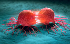
Researchers use different techniques to show that a cell is more than just the sum of its parts.
|
The isolated parts of a cell can only tell you so much. To see what’s really going on, you need to look at the whole cell.
Experimentalists have always reveled in taking things apart. And, over the years, this has led to profound understanding—of the parts. The push now is to put things, such as the cell, back together, to query the entirety of the system. As recently reported in the Journal of Biomolecular Screening, Paul Lee, PhD, principal scientist, Amgen, Thousand Oaks, Calif., has found one such way to look at membrane-bound G-protein-coupled receptors.
“For the last 30 to 40 years, there has been a gold standard for this ligand-receptor binding assay,” says Lee, “but the problem is that it’s not homogeneous.” After being exposed to the ligand, the membrane is run through multiple washing steps, thereby making it impossible to get any sense of equilibrium binding. “You also have to [lyse] the cell membrane expressing the receptor of interest, so we’ve disrupted the native environment. It’s very hard to do any real-time, kinetic measurement in that format,” he explains.
Lee worked on this problem before, but lacked the proper optical technology to solve it. “In the past, you have to use a high-powered instrument in order to focus on the bottom layer—and those instruments are very expensive,” and data collection is both piecemeal and time-consuming. “But this new instrument (IsoCyte, a benchtop laser scanning cytometer, from Blueshift Biotechnologies, in Sunnyvale, Calif.) has an optical path, which allows you to scan the whole plate, while still allowing you to capture a monolayer of cells at the bottom of the well,” Lee says.
To validate this method, Lee used a Cy3B-labeled telenzepine fluorescent ligand, and CHO cells expressing M1 muscarinic acetylcholine receptors. Without any washing or separation steps, he was able to capture real-time binding kinetics data using a relatively small sample size. “This is proof of the generalized method. In the future, if anyone’s interested in cell surface receptors, they won’t have to grow liters of cells, break down the cells, get the membrane. … you can already generate data (from the well). And in the whole cell environment, the data will be far more robust,” he adds.
Brain storm
Watching a whole cell go about its household chores is one thing, watching it differentiate, and discovering how to induce it is quite another. “Our in vitro assay system is built around looking at the differentiation of human neural stem cells,” says Todd Carter, PhD, director of biology at the San Diego, Calif.-based BrainCells, Inc. “What we’re interested in is trying to identify compounds that modulate adult hippocampal neurogenesis.” There are other groups doing this sort of work, but Carter asserts that his is the first cell-based platform to use human neural stem cells for the purpose of drug discovery.
The first step was to create the appropriate cell lines; for genetic reasons, ready-made sources wouldn’t do. After deriving stable, multiply-independent neural stem cell lines, the BrainCells team created the assay. “It was very challenging to develop, [i.e.] translating a primarily low throughput activity—looking at a slide under a microscope—into something that can be quantified out of a 96-well format.”
Carter next had to contend with the inadequacy of known reporter constructs that failed to be predictive of neural differentiation, relying instead on a combination of morphologic changes and the presence of protein markers. The system also had to function over time, as this is not a single-day assay.
Having ironed all that out, BrainCells began screening known compounds for neurogenic activity. That said, the end-purpose may elude a simple guess: Carter is looking for drugs to treat depression. “There are already antidepressant drugs that effect neurogenesis, but nobody ever looked at it that way before. One of our founders, Dr. René Hen, showed that neurogenesis and depression are linked. He showed that all marketed antidepressants increased neurogenesis in rodents,” Carter says. It was further discovered that in humans, the two- to four-week lag time in therapeutic response to antidepressants corresponds to the time it takes for human neurogenesis to occur—the behavioral and morphologic changes are linked.
|
It ain’t chopped liver
Brain function is not a measure of one type of brain cell, and a liver is not just hepatocytes. Dawn Applegate, PhD, president and CEO of RegeneMed, San Diego, puts it this way: “Think of it as raisin bread. The raisins are the cells and the bread is the tissue they create. The cells have the capability of making all the tissue components, all the extracellular matrix proteins, all the growth factors …” Applegate and colleagues are proponents of a system that recreates the entire loaf, with this occurring through the creation of 3D liver tissue cultures.
Toxicity assays have, to date, used cellular fractions, recombinant enzymes, or primary cell cultures in a dish, but Applegate considers these meager substitutes for a complex system. “If you’ve lost the native architecture, you’ve lost physiologic relevance. It’s not a true mimic of what’s going on in the body.” RegeneMed’s matrix is not, however, to be confused with culturing in 3D gels. Yes, cells will take on a rounded morphology, “but it’s like someone swimming in sea of molasses,” says Applegate. “What sort of useful work can they do?” Recent studies at Johns Hopkins University (Baltimore, Md.) have shown that a RegeneMed-type environment allows for cells to rotate, translate, and co-migrate with neighboring cells, thereby enhancing tissue-specific function.
The cultures are also particularly long-lived, and this, as capstone to their experimental utility, has attracted a great deal of interest from industry. “A lot of the pharma companies are doing side-by-side testing of all of the 3D assays that are out there, trying to pick the best one for their toxicologic needs.”
On the horizon
There is perhaps no greater need for cell-based assays than in the field of oncology, where one cancer does not fit all. Consider the NCI-60 panel—a resource with wide genomic variations. “Ideally, you want a system where the differences in cell lines are minimized, so that any effects you see can be traced to a specific mutation that you’ve introduced,” says Rob Howes, PhD, R&D Director, of Horizon Discovery, Cambridge, UK.
In the age of targeted therapies, it’s essential to know just where the effect is felt.
A frustration with state-of-the-art models is what drove Howes to co-pioneer a system whereby singular, specific oncogenic mutations can be reliably introduced into otherwise normal, immortalized cells lines. “The new technology uses a viral packaging system,” explains Howes. “Take a plasmid with the gene-of-interest and pass it through a viral packaging system and, for some reason … it produces a recombination rate of up to 25%.” This efficiency far greater than previously-known techniques raises the technique from an academic exercise to something that can be commercially-viable.
The technique is also applicable across multiple cancer types. For example, Horizon currently uses a breast tissue cell line, yet Howes can introduce mutations that translate into the behavior of a cancerous colon cell; as long as the prominent oncogenic changes of a given cancer are known, the derivation of the parental line is not an issue.
“This is a great way to test if your drug is sensitive to cells that harbor those mutations,” says Howe. “It even allows you to target un-druggable drug targets, say K-Ras or p53. As long as you see selectivity in mutant vs. parental, then you don’t initially care where the drug actually goes to because you see can the effect on activity.”
And there is growing activity around Howes’ technology, commercially-known as GENESIS. Genentech, Novartis, and Millennium: The Takeda Oncology Company are part of a growing client list.
About the Author
Neil Canavan is a freelance journalist of science and medicine based in New York.
This article was published in Drug Discovery & Development magazine: Vol. 12, No. 1, January, 2009, pp. 24-27.
Filed Under: Drug Discovery


