Membrane proteins are difficult to purify, but wizards of protein purification may send this process on a one-way trip to easy-land.
 click to enlarge  Schematic drawing of an Affinity Grid (top), which features a dried lipid monolayer containing Ni-NTA lipids. His-tagged membrane proteins will specifically adsorb to the Ni-NTA lipids on the Affinity Grid, which can then be visualized in the electron microscope (bottom). (Source: Tom Walz, PhD) |
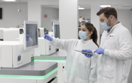
Protein purification is probably one of the most challenging endeavors a life scientist can pursue. It is not for the faint-of-heart. And many failures lurk behind every success story. Now, throw high levels of hydrophobicity and poor solubility into the mix and the failure rate increases exponentially with every attempt. That is the uphill battle faced by the brave souls who attempt to purify membrane proteins. However, like Dorothy sang in the classic American film The Wizard of Oz: wishes do come true “somewhere, over the rainbow.” And with the tools for membrane protein purification described in this article, some wishes might come true.
In his very technology-oriented lab, Ole Nørregaard Jensen, professor, Department of Biochemistry and Molecular Biology, University of Southern Denmark, Odense, spends much of his time developing mass spectrometry (MS)-based tools to study posttranslational modification of membrane proteins. Such methods are devised to study everything from the single membrane protein to the membrane proteome. But MS is at the end of this process. And the first steps in the process depend on the type of protein isolated. For example, many receptor proteins such as the epidermal growth factor receptor (EGFR) must be immunoprecipitated following release from the plasma membrane. This involves a protocol starting off with on the order of 107 cells. The protein is immunoprecipitated, followed by isolation by gel electrophoresis. Trypsin digestion of the isolated protein into peptides prepares it for liquid chromatography, which is coupled with tandem mass spectrometers. Data are interpreted using the MASCOT search engine. This is an example of how they study a single membrane protein using a classical technique.
 click to enlarge The one-day protocol for membrane protein expression and purification using the MembraneMax Protein Expression Kit. (Source: Invitrogen Corporation) |
Jensen uses a cell line containing an abundance of EGFR, making its purification easier than most membrane proteins he currently studies. However, the solubility of other membrane proteins presents a challenge to their purification. “And also many of them are heavily modified by glycosylation and other modifications so they can be difficult not only to extract but also to digest into peptides,” says Jensen. “Sometimes it can be difficult to digest these proteins if they are very hydrophobic and have many transmembrane regions.” To overcome this digestion problem, Jensen and his colleagues are trying to optimize detergent extraction conditions by using different detergents and trying to optimize digestion conditions for the highly hydrophobic membrane proteins by using, for example, an organic solvent in the digestion buffer. “We start out with the classical protocols and then we adapt them to work with MS. So if the classical protocol calls for the use of detergents that are incompatible with MS, we need to tweak the protocol to make them work for our purpose. … I think for proteomics it is worthwhile to modify the established protocol to avoid bias caused by certain solubilization conditions.”
A sweet approach
Jenson and colleagues are also investigating the plasma membrane proteome of stem cells to gain a better understanding of how these cells differentiate. In this case, the crude cell lysate is first separated by centrifugation in a sucrose or Ficol gradient to remove all of the organelles and purify the plasma membrane. The crude plasma membrane fractions are then washed with sodium carbonate to remove loosely-attached proteins. In the end, the plasma proteome is digested with proteinase Lys C, then with trypsin, and finally identified by MS, with additional phosphorylation site mapping, also done here by MS.
Another researcher who has used the sucrose gradient to purify membranes is Weiqing Zeng, PhD, visiting research associate, DOE-Plant Research Laboratory, Michigan State University, East Lansing. Zeng, who studies enzymes involved in plant cell wall biosynthesis, is interested in determining the locations and topologies of these enzymes in plant cells. “To [determine topology], you use a combination of the antibodies against your own protein and also use proteases that degrade just your protein. And by playing with the digestion of just your protein at different locations, you will see which part of your protein is located either on the outside of the membrane or facing the lumen of the membrane fraction,” says Zeng. Following separation of the different membranes isolated from plant cells or tissues, Zeng assays each membrane fraction for activities of these biosynthetic enzymes of interest and for the presence of marker enzymes that identify the type of membrane present.
|
Affinity for metal
QIAGEN develops kits for the pharma and biotech industries, as well as for academic researchers. So they don’t typically work with one protein but rather with several bacterial and human natural and synthetic genes at a time. They work with a number of different proteins expressed in vivo and in vitro in bacteria, insect, and mammalian cells. Their newest kit in development, called the Nickel-NTA membrane protein isolation kit, is scheduled to hit the market in January 2009. Here’s how the kit works. The first step in this process is to express the protein in cells. And in the case of this kit, the protein must be histidine (His)-tagged.
Once the protein is expressed and present on a cellular membrane, the kit allows the user to try seven different carefully-selected detergents to determine the best solubilization conditions for their protein. Users then perform a Western Blot analysis to determine how much of the total target membrane protein could be solubilized with each detergent. “After solubilization, it’s possible to purify the membrane protein with the most suitable detergent in the kit,” says Jan Kubicek, PhD, R&D scientist, at QIAGEN GmbH, Hilden, Germany. The protein is purified on the Nickel-NTA column, which works on the principle of metal affinity chromatography and is provided with the kit. After the protein is loaded onto the column, it works just like a normal chromatography protocol, i.e., wash, elute, measure, and visualize via SDS-PAGE. “We created a kit to make this really difficult field work easier for scientists.”
Thomas Walz, PhD, professor and Howard Hughes Investigator, Department of Cell Biology, Harvard Medical School, Boston, Mass., created an electron microscopy grid that works on the same principle as the Nickel affinity chromatography column. It is called the Affinity Grid and it is a carbon film, on top of which sits a lipid monolayer containing lipids modified with a Nickel affinity group. Walz started developing this method around 18 months ago, with the goal of creating a technology that allows researchers to visualize the structure of any His-tagged protein without the need for purification. The basic protocol involves expression of the His-tagged protein in the desired cells and then lysing those cells followed by centrifugation. For soluble proteins, the cytosolic fraction is applied to the Affinity Grid. For membrane proteins, the membrane fraction is detergent-solubilized and applied to the Affinity Grid. The grid with the specifically bound His-tagged proteins can then be stained or frozen and inspected in the electron microscope. The Affinity Grid was originally designed for the isolation of protein complexes as these are typically unstable and tend to disassemble during long purification procedures, but it is equally useful for the visualization of membrane proteins. “We developed [the grid] with the hope that other people would use it. So we published on it and we hope that a company is going to produce the grid,” says Walz.
Purification for membrane proteins may be difficult, but new tools for expression, purification, and analysis will likely make this process easier in the future.
This article was published in Drug Discovery & Development magazine: Vol. 10, No. 9, September, 2008, pp. 32-34.
Filed Under: Genomics/Proteomics