
Introducing Deepcell’s REM-I, which applies AI to cell morphology analysis.
From her early days as a research assistant at UCLA, to her work as a postdoctoral fellow at Stanford, Deepcell co-founder and CEO Maddison Mahdokht Masaeli has actively engaged in the field of biomedical engineering. Now, Masaeli and her team at Deepcell, a company she co-founded in 2017, are introducing a new strategy to drug discovery with the launch of REM-I. The platform combines AI and morphological analysis, a technique that studies the form and structure of organisms and their specific structural features.
“The REM-I platform offers several unique capabilities for deep assessment of single cell images and the ability to sort cells without bias,” Masaeli explained in an interview. “It is specifically designed to expose and analyze the heterogeneity within samples, as well as the heterogeneity in response to therapies, all at single cell resolution.”
A core feature of the REM-I platform is “its ability to provide a comprehensive characterization and sorting of viable cells,” Masaeli noted. This capability gives researchers the ability to assess the viability and overall health of individual cells within a sample. “The platform can identify any morphological changes that occur when exposed to different therapies and sort subpopulations of cells that are responding or resisting to the therapy,” she added.
Such detailed analysis from the REM-I platform could streamline the identification process for potential drug candidates, helping support researchers’ understanding of how such candidates interact with cellular structures and functions.
Decoding cellular morphology with the Human Foundation Model
Deepcell’s Human Foundation Model (HFM) is an AI model that has been trained using millions of cell images. By not predefining any specific features or structures to look for in the cells, the AI model is allowed to discover and learn important features from the data itself, a process known as unsupervised learning. The proprietary HFM takes a thorough approach to hypothesis generation and testing, extracting visual features from a large and diverse set of cell images in a self-supervised fashion. “We have validated the model over a wide range of both synthetic beads and cell types with impressive performance,” Masaeli said.
HFM enables the prediction of a therapeutic agent’s potential impact on cellular structures and functions, leading to an improved understanding of its mechanism of action, efficacy and potential side effects.
Deepcell uses AI-driven approach for high-dimensional cell biology
Menlo Park-based Deepcell’s approach involves training the AI model on an informationally diverse subset of 2 billion single-cell images. Thie company used unlabelled training to allow the generation of deep learning embeddings and morphometric analysis in high-dimensional vector space.

Maddison Mahdokht Masaeli
“We’ve amassed 2 billion single-cell images and selected an informationally diverse subset to train the Human Foundation Model,” Masaeli said. “The unlabelled training process allows us to generate deep learning embeddings and morphometric analysis in a high-dimensional vector space.” Masaeli is convinced that this approach could open up novel avenues in biology that can’t be captured through genomics, transcriptomics or other highly quantitative biomarkers.
Real-world applications of REM-I
In the recent Series B funding round, Deepcell raised $73 million to further advance its AI-powered single-cell analysis platform. Koch Disruptive Technologies led the funding round. New investors joining the round included Bridger Healthcare, Horizons Ventures, Casdin Capital. Also contributing were lead investors from previous rounds, Andreessen Horowitz and Bow Capital.
Prominent individuals such as Jeff Dean, head of Google Brain, and Matt Mcllwain, managing director at Madrona Venture Group, also took part. “Since the inception of the company, we have been fortunate to be backed by top investors who combine domain expertise and exposure with extensive network and long-term perspective,” Masaeli said.
Masaeli believes that the current applications of AI-powered morphology, including the ability to uncover patterns not readily captured through genomics, transcriptomics, or other highly quantitative biomarkers, mark a new beginning of what the technology can achieve.
Quantifying cell morphology at scale
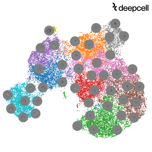
Earlier this year, Deepcell released data sets to enable researchers to explore high-dimensional morphology data.
Deepcell’s approach to quantify and scale morphological assessment through standardized high-resolution imaging supports the integration of this phenotype into the multi-omic understanding of biological samples. “In cancer biology, this means improving our ability to deconvolve complexities of these samples, And an approach to purify certain cells of interest, even the most fragile ones, for further assessment,” Masaeli added. “In gene therapy, the product can be used to characterize cell phenotype in a quantitative and high-dimensional manner.”
Deepcell’s Technology Access Program helps validate the product’s performance in real-world settings and facilitating collaborations with top researchers. “The results of some of these studies are in the process of publication for public access,” Masaeli said.
Maseli notes that the company is partnering with major research institutions internationally to provide them with early access to its products. “Our partners have used the technology for a variety of research areas, including liquid biopsy, drug screening, cancer biology and CRISPR screening,” she said. “The results of some of these studies are in the process of publication for public access.”
Filed Under: Cell & gene therapy, Data science, Women in Pharma and Biotech





Tell Us What You Think!
You must be logged in to post a comment.